Minimally Invasive Spine Surgery
Lumbar Discectomy/ Spine Fusion, Interbody
Fusion/Herniated Disc
When nerves in the lumbar spine are pinched, as may
occur in the case of a herniated disc or lumbar spinal stenosis, pain may
radiate down the lower extremities. This is known as lumbar
radiculopathy. In more extreme cases, weakness of
the lower extremities may occur, and in more critical conditions, a patient
may lose bowel and bladder control. Sometimes, all that is needed is a discectomy
surgery (minimally invasive surgery), which
involves removal of the herniated
lumbar disc, and some of the lumbar
disc material between the vertebral bodies. At other times, a lumbar
decompressive laminectomy surgery (minimally invasive surgery) is required for a broader decompression of the nerves. At
times, the spine may become unstable, either preoperatively as a result of
injury or degeneration, or during back surgery, as a result of the extensive bone
removal which is sometimes required.
We will discuss back surgery (minimally invasive spine
surgery) which consists of a
lumbar discectomy or lumbar laminectomy to relieve radiculopathy. We
will also cover surgery for lumbar spine fusion.
Much of this will be done through a minimally invasive approach.
(Images were provided by Medtronic, Inc.)
When performing a minimally invasive spine surgery (back surgery), such as a discectomy,
laminectomy, spine fusion, or minimally invasive surgery, the patient is
lying on their abdomen, supported in a variety of ways. The patient
may be positioned on gel rolls, or on a Wilson frame, or on an OSI or
Jackson table, which are designed specifically for surgery on the spine.
For lumbar
surgery, an incision is made over the lumbar spine. The exact position
and length of the incision depends upon what needs to be done. After
the incision is made, the muscles are pulled away from the spine.
Below, we see an open back (spine) surgery, with retractors in place, pulling muscle and
fat (adipose tissue) to the side, so that the lumbar spine is exposed.
An instrument is used to help
with the removal of the lumbar facet.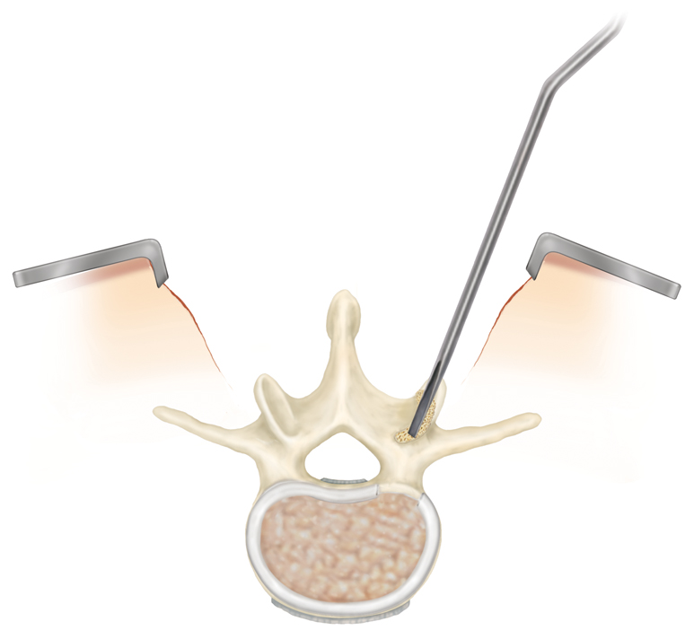
A tubular dilator is passed down to the lumbar spine for the minimally invasive surgery
on lumbar the spine. The bottom portion of the tubular retractor is docked
on the lumbar lamina.
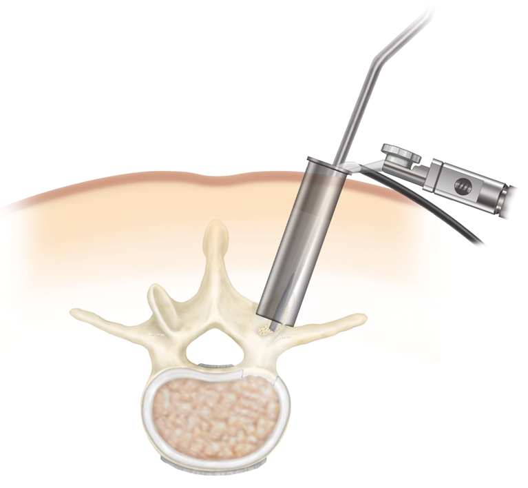
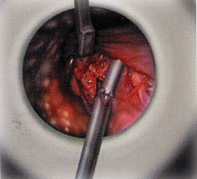
Below is the view which the surgeon has (during a
minimally invasive spine surgery), with the lumbar nerve root seen. The
surgeon is performing a discectomy, which is a removal of the lumbar disc.
The view is what the surgeon would see while looking and working through the
tube.
Below, a lumbar disc distractor is placed within the
disc space. This helps to maintain the height of the lumbar disc space
on one side, while the other side is being prepared.
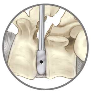 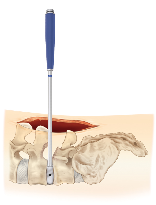
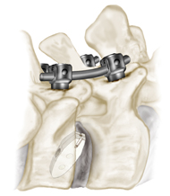
Pedicle screws may be placed within the pedicles of
the lumbar spine, and on the opposite side, to help create a working
channel. Pedicle screws are screws which go into the pedicles, and
provide a rigid and stable construct to which rods may be attached.
The rod may then be attached to a pedicle screw placed at another spinal
level.
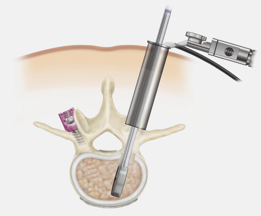
A rotating cutting device is placed within the disc
space to clean out some of the lumbar disc material.
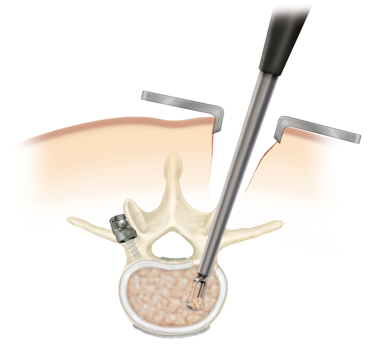
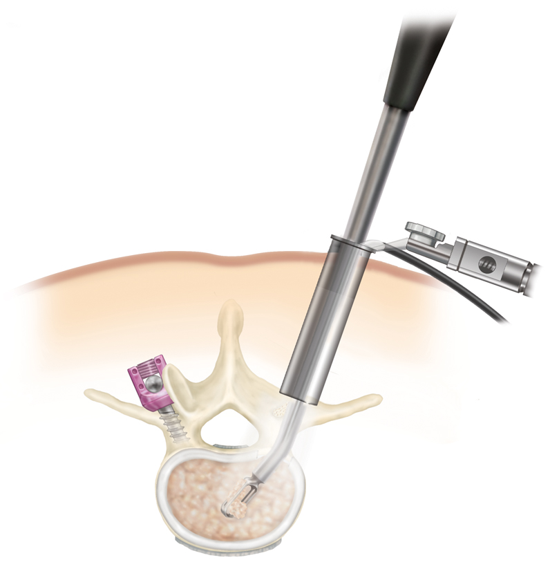
Curettes are used to scrape the endplates of the
vertebral bodies, further cleaning out the disc space.
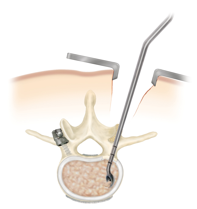 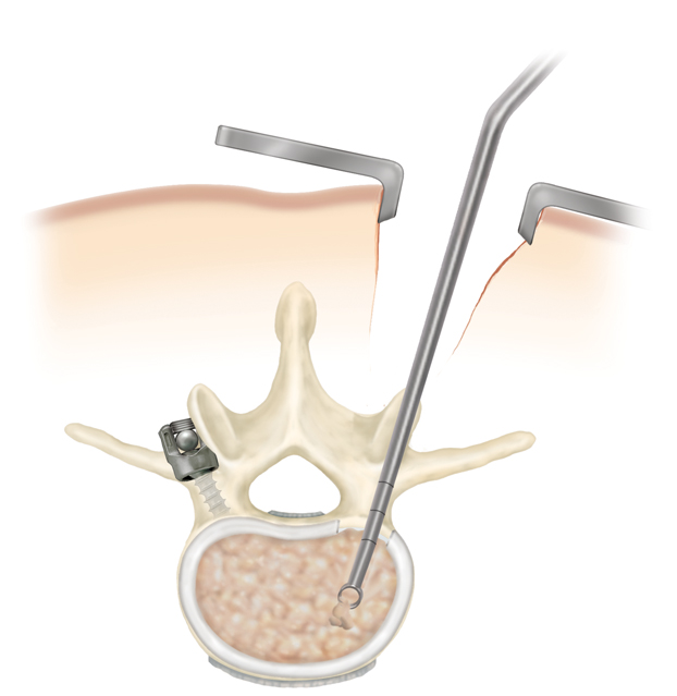 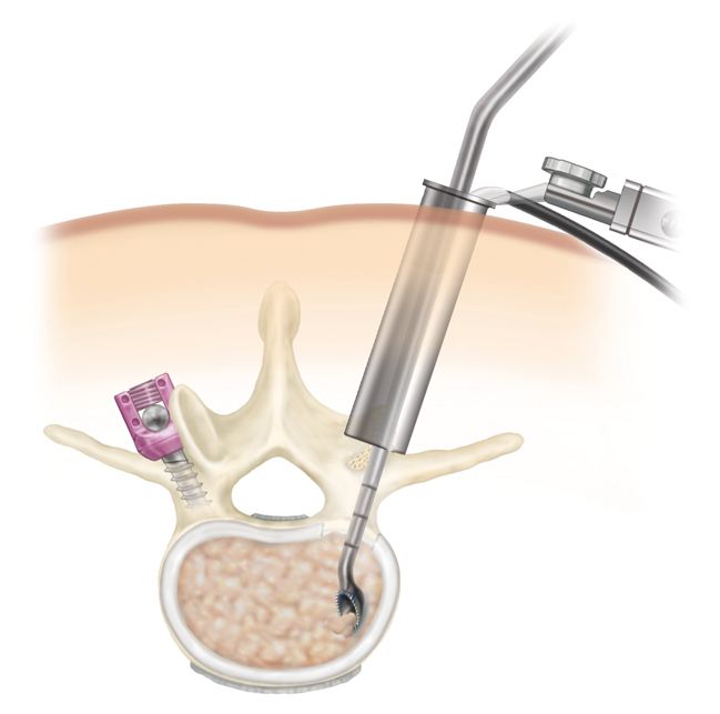
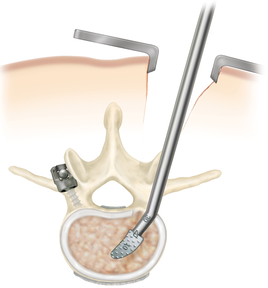
A rasp is used to roughen up the edges of the bone in
the vertebral body, which will place the eventual intervertebral body graft
in contact with healthy bleeding vertebral body. Below, on the left, this is
done through an open insicion, whereas on the right, it is done through a
minimally invasive approach tubular guide. On the left, the spine
surgery is being accomplished through a Wiltse approach. The Wiltse
approach involves an incision off of the midline, with dissection through
the various muscle layers of the back.
 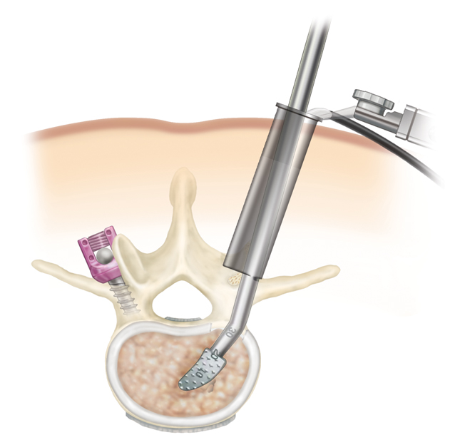
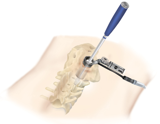
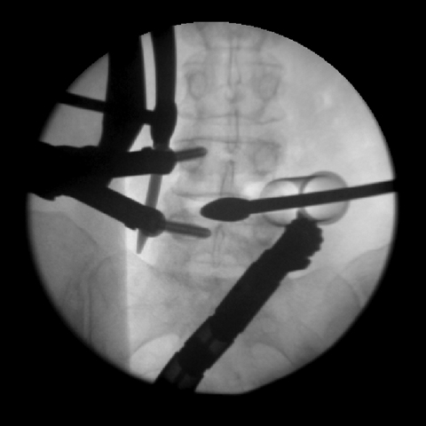
Below, we see a minimally invasive retractor in place.
On the right is an x ray of a disc distractor in place, and pedicle screws
and a rod are seen.
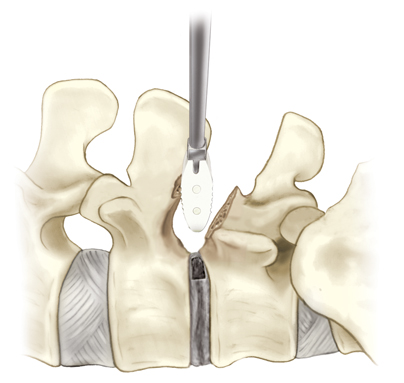
Below, a trial is performed to find the appropriate
sized intervertebral body implant. This allows the surgeon to find the
appropriate graft for the placement of a posterior lumbar interbody fusion (PLIF).
A PLIF is a graft placed between the vertebral bodies, which will allow one
body to fuse to the adjacent one.
 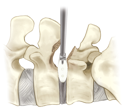 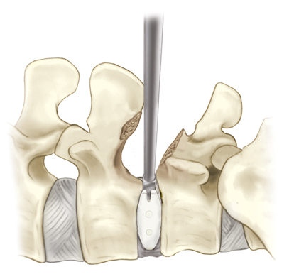
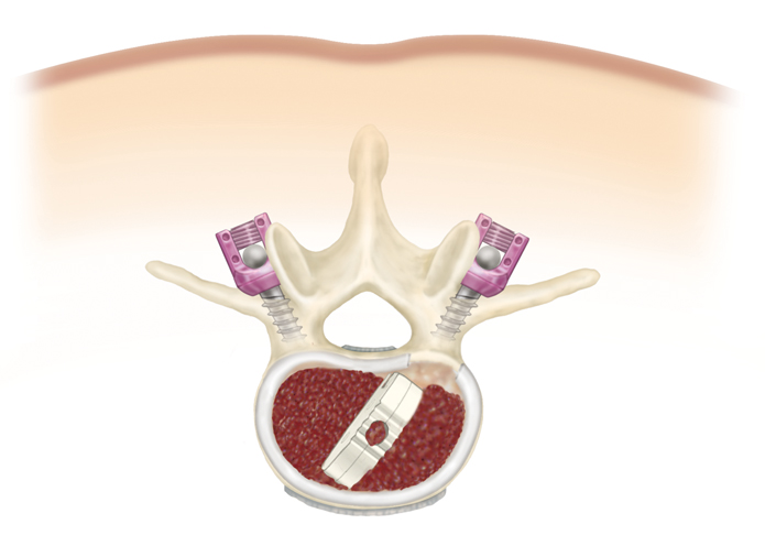
Next, the intervertebral cage is deployed into the
intervertebral disc space. Bone graft is packed around the cage.
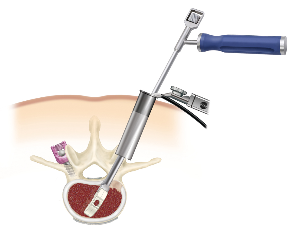
Pedicle screws are placed on both of the lumbar spine,
to assist with the spine fusion.
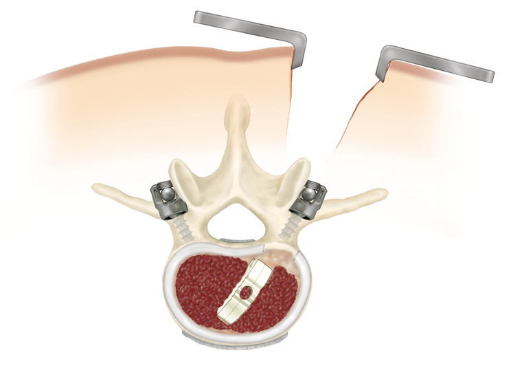
Pedicle screws and rods are placed on both sides, to
promote lumbar spine fusion.
|

