interesting cases
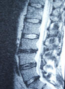
Patient AY is a 36 year old man, who has had a 5 year
history of significant low back pain. He has no lower extremity pain.
His weight is 360 pounds, and height is 5'5". His physical exam was
significant for tenderness to palpation over the lower lumbar area.
There was no weakness in the lower extremities. MRI scan of the lumbar
spine, seen here, is notable for degeneration of the three lower disk spaces
(the disk spaces which are normally white on this MRI sequence, are instead
dark).
Treatment strategy: Although there is
degeneration of the disk spaces in the lumbar spine, this patient was
handled in a conservative manner. Initially a weight loss and exercise
program were implemented. The patient still has pain, although it is
improving. If, after substantial weight loss, the pain persists, then
we will have the patient undergo a discogram, and if indicated, consider an
anterior lumbar interbody fusion (ALIF).
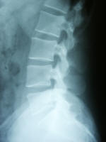 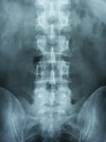
Patient TK is a 28 year old man who has had 5 weeks of
severe low back pain an spasm. There is no pain in the lower
extremities. Neurological examination is intact. The lumbar spine x
rays are normal. This patient responded very well to conservative
care, which included conservative care, stretches, and massage. Pain
resolved completely.
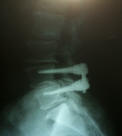
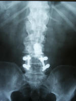
Patient RT is a 56 year old man, who had a four year history of significant
pain radiating to the lower extremities, along with low back pain. MRI
scans of the lumbar spine showed significant spinal stenosis at L4/5.
He underwent a decompressive laminectomy at L4/5, and a fusion with pedicle
screws and rods, and a bone growth stimulator. Postoperatively, he did
very well, with significant improvement in low back and lower extremity
pain. On the x-rays, the pedicle screws and rods can be easily seen in
the lateral lumbar x-ray (left) as well as the AP x-ray (right).
  Patient
CM is a 19 year old woman, who has had a 6 month history of worsening
thoracic and lumbar pain. She has no pain radiating to the lower
extremities, and there is no difficulty with bowel or bladder control.
Neurological exam was completely normal. Patient
CM is a 19 year old woman, who has had a 6 month history of worsening
thoracic and lumbar pain. She has no pain radiating to the lower
extremities, and there is no difficulty with bowel or bladder control.
Neurological exam was completely normal.
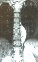
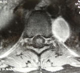
On MRI she was found to have a tumor adjacent to the
thoracic spine. It is seen on the axial cuts (above left) as a bright
ovoid mass adjacent to the spine, and on the coronal images (above, right)
again as a sausage shaped mass adherent to the spine. She
underwent a transthoracic gross total removal, and has had a good recovery.
She suffers no lower extremity pain, mild low back pain, and her returned to
work.
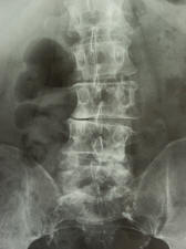
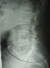 TJ is a
65 year old man, who has had 5 years of low back
pain, and two years of lower extremity pain.
The pain radiates to both legs when walking and
standing, but improved with sitting. We can
see from the plain x rays that there is a degree of scoliosis. On the
sagittal TJ is a
65 year old man, who has had 5 years of low back
pain, and two years of lower extremity pain.
The pain radiates to both legs when walking and
standing, but improved with sitting. We can
see from the plain x rays that there is a degree of scoliosis. On the
sagittal
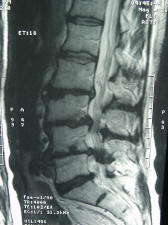 MRI
we see that there is significant stenosis of the lumbar spine. There is also
a grade I spondylolysthesis (slip) of L3 on L4. This patient underwent
a lumbar decompression, but at surgery, the bone was too weak and
osteoporotic to adequately hold pedicle screws. The patient did
well, and had significantly improved lower extremity function, although a
mild amount of low back pain persisted. MRI
we see that there is significant stenosis of the lumbar spine. There is also
a grade I spondylolysthesis (slip) of L3 on L4. This patient underwent
a lumbar decompression, but at surgery, the bone was too weak and
osteoporotic to adequately hold pedicle screws. The patient did
well, and had significantly improved lower extremity function, although a
mild amount of low back pain persisted.
|

