|
The arrival at a diagnosis, in medicine, requires a history, physical exam,
and tests. Generally, 85% of the diagnosis relies upon the history.
When it comes to low back and leg pain, important questions include:
- when did the pain begin
- what precipitated it; was there an injury, or did it occur
spontaneousluy?
- does it stay in the back, or does it travel down the leg, and it so,
where in the leg does it go
- what makes the pain better, and what makes it worse
- is there any weakness associated with it
- is there any loss of bowel or bladder control
Next, the physical exam should contribute 10 % to the diagnosis. During
the exam, specific things the physician will watch for are:
- tenderness to palpation over the lumbar spine
- signs of "straight leg raising" or "crossed straight leg raising":
a straight leg raise is a test in which the leg is raised straight up in the
air, extending the knee. If pain shoots down the leg being raised,
this is a positive indication that there is likely to be something (quite
possibly a herniated disk) pushing on the nerve root. A crossed
straight leg raise test is a test where, when one leg is raised, the pain
travels down the opposite leg. This sign often has a 96% accuracy in
predicting the presence of a herniated disk.
- weakness in the lower extremities. The lumbar and sacral nerve
roots each supply different muscle groups. Pressure on a nerve root
will often cause weakness in the muscle supplied by that root. For
example, the S1 root supplies the gastrocnemius muscle (calf muscle), and
injury to or pressure upon the root may cause the patient to have difficulty
walking on the toes. Pressure or injury to the L5 root may cause
difficulty picking up the big toe, and can result in foot drop.
- sensory changes in the lower extremity, in the distribution of the nerve
being affected. The S1 root supplies sensation the the lateral aspect
of the foot, and injury to the root can result in numbness to the lateral
foot and and little toe.
- reflex changes can also result from pressure on a nerve. The S1
root is responsible for the achilles reflect, and injury to this root may
result in an absent ankle jerk.
Finally, we tests are performed. Typical tests a patient with low back
and lower extremity pain may undergo, include lumbar spine x-rays (films), CT
scans (computed axial tomography scans), MRI scans (magnetic resonance imaging),
myelograms, post myelographic CT scans, EMG/NCV (electromyogram/nerve conduction velocity) studies, discograms,
and bone density tests.
Lets go over the value of each.
- lumbar spine films: these plain x-rays are good at showing
alignment of the spine. Any slippage, known as spondylolisthesis or
subluxation, is easily visible on lateral x-rays. Also, compression
fractures of the vertebral bodies is easily seen. Tumors eating away at
the bony structure of the spine can also be detected, as the done defect (or
lytic lesion) is often seen.
- CT scans: these show "cross sections" of the spine. This can
be thought of as slices of bread. If we have something within the bread,
perhaps a sausage, taking a picture of the bread won't show the sausage.
But, if we slice the bread, with each slice being 5 millimeters thick, and if
we look at each slice laying down on its side, we will now be able to
accurately figure out what is buried within the bread. The same holds
true for CT scans. They are "bread slices" of the body, accurately
revealing the anatomy within. When used on the spine, they detail bone
very well, but are not quite as good at showing soft tissue, such as herniated
disks, nerves and tumors. CT scans use x-rays, which are send through
the area of interest in numerous directions, anud then a computer adds the
images in 3-dimensions, to "calculate beam attenuation," and then display this
in a manner, with pictures, that a human can understand.
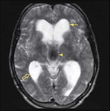
- MRI scans: the images provided are similar to CT scans, in as far
as these images provide serial slices through the lumbar spine, or other areas
of interest. They use magnetism, instead of x-rays, to abtain the
images. They can also provide images in other planes, beside the axial.
These other planes are sagittal and coronal. Of interest, it is easy to
remember what direction these other planes describe if one thinks about where
the names were derived from. A sagittal slice is a vertical slice
through an object, and it comes from the vertical slice made by Sagittarius
and his bow and arrow. A coronal slice describes a slice taken from side
to side, and comes from the slice made by the crown a queen would wear, from
side to side on the hear (corona means crown).
Besides showing information in additional planes, the MRI gives much better
detail of the soft tissue anatomy of the spine, than does CT. Disks,
nerve roots, and tumors are all seen much better. On the other hand,
bone is seen better on CT scans.
- myelograms: a myelogram is a study in which the radiologist
performs a spinal tap, places a radioopaque dye (a dye which shows up on x-ray
and CT scans) into the spinal fluid, and then x-rays and post myelographic CT
scans are performed. What is provided is an "outline" or "shadow" of the
pathology. For example, instead of seeing a nerve root sheath filling
nicely from the dye in it, it may show nice filling to a point, and then show
a bump in it. These studies are often performed in patients where the
MRI studies are ambiguous, or in patients who have a pacemaker implanted for
the heart ( pacemakers cannot be used in the presence of an MRI machine).
Although they have been used very frequently in the past, MRI scans are
obtained much more commonly now because they are non invasive. Nobody
likes to get a needle stick if they don't have to.
- post myelographic CT scans: with the dye in the spinal canal,
w-rays can show the pathology of nerve root compression, but CT scans can show
it with much more detail.
- EMG/NCV studies: these studies monitor the electrical functioning
of nerves and muscles. The other studies mentioned monitor appearance or
anatomy, but this study looks at function. There is a difference.
Imaging taking a picture of a person. You could tell how tall he/she
was, and what color hair they had. But you would be unable to determine
whether they had diabetes or high blood pressure. A blood sugar test or
a blood pressure measurement would tell that. They are tests of
function. In a similar way, an EMG/NCV is a test of function, looking at
how well a nerve works.
- Discogram: A discogram is a test which looks at function and form
(anatomy). During the test, a needle is placed within the disk space,
and dye is injected under pressure. If this pressure injection of dye
causes low back pain which is the same as the low back pain a patient
typically experiences, this signifies that some of the low back pain may be
coming from degeneration or micro-instability within the disk space, and the
patient may potentially benefit from a fusion. In addition, x-rays and
CT scans are taken with dye in the disk spaced, and information is gathered
about tears within the annulus of the wall of the nucleus polposus.
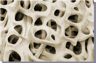
- Bone density tests: this test measures the density and strength of
bones. Osteoporosis is the leading cause of vertebral compression
fractures. A number of tests are available, but the most common is the
DEXA (dual energy x-ray absorptiometry) test.
|