|


last
updated
June 19, 2009
 
................................
Other areas of
Expertise:
Gamma Knife
Normal Pressure-
Hydrocephalus

Osteoporosis: Prevent/Reverse It

Trigeminal Neuralgia

| |
Lumbar Discectomy
Indications for surgery
- pain radiating to lower extremity in distribution
of a nerve, which does not improve with 6 weeks of conservative care
- patient reluctance to wait for 6 weeks of
conservative care, especially if it is likely they will need surgery in
any case
- progressive motor deficit or
cauda equina syndrome.
Surgery
 Lumbar discectomy involves the removal of a small
amount of the roof of the spinal canal, known as the
lamina. The operation is typically done under magnification,
with the surgeon using either an operating microscope or loupes (small
magnifying glasses worn by the surgeon, like a pair of glasses). The
nerve roots are gently moved aside, and the herniated disc removed (in the
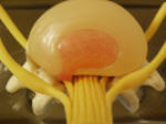
illustration on the right, a small herniated disk (red) is
seen pushing into the nerve roots (yellow)). During a typical discectomy, roughly 75% of the disk in the interspace is removed as well.
Then, the would is closed. Lumbar discectomy involves the removal of a small
amount of the roof of the spinal canal, known as the
lamina. The operation is typically done under magnification,
with the surgeon using either an operating microscope or loupes (small
magnifying glasses worn by the surgeon, like a pair of glasses). The
nerve roots are gently moved aside, and the herniated disc removed (in the
illustration on the right, a small herniated disk (red) is
seen pushing into the nerve roots (yellow)). During a typical discectomy, roughly 75% of the disk in the interspace is removed as well.
Then, the would is closed.
Sometimes a discectomy is referred to as a
laminectomy, because a small amount of the lamina, or roof of the spinal
canal, may be removed in order to get to the herniated disc and disc
space.
Outcomes
85-90% of appropriately selected patients have good
outcomes, with improved pain. Leg pain is improved more
consistently than low back pain.
Complications
superficial wound infection: 2%
deep wound infection: 1%
increased motor deficit: 1-8%
dural tear (resulting in spinal fluid leak): 5%
recurrence of disk herniation: 6%
|
|

